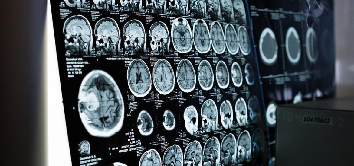
A team of researchers from the Institute of Neurosciences CSIC-UMH in Alicante, Spain, managed to obtain images of brain inflammation using diffusion-weighted magnetic resonance, according to a study published in the journal Science Advances. The authors believe this approach could become a valuable tool for diagnosing conditions like dementia and other neurodegenerative diseases.
This method of obtaining images from the brain is similar to a conventional MRI, but it needs data acquisition sequences and unique mathematical models. Using this system, the team was able to quantify alterations in the brain caused by changes in different types of nerve cells that are involved in the inflammatory process in the brain.
Degenerative diseases like Alzheimer’s or multiple sclerosis can be challenging to diagnose. Researchers know that activation of two types of brain cells — microglia and astrocytes — are involved in the inflammatory response in the brain and contribute to disease progression, but this mechanism is difficult to assess noninvasively in patients. It is possible to use positron emission tomography (PET), but this approach exposes patients to ionising radiation, limiting its use to vulnerable populations and some clinical studies. In addition, PET has a low spatial resolution, making it unsuitable for detecting small changes occurring in different cell types.
In contrast, diffusion-weighted MRI can image brain microstructure noninvasively and with high resolution by capturing the movement of water molecules in the brain, which then generates contrast in MRI images. Using this approach, the Spanish researchers developed a way to “see” activation of microglial and astrocyte activation during inflammation.
“This is the first time it has been shown that the signal from this type of MRI can detect microglial and astrocyte activation, with specific footprints for each cell population. This strategy we have used reflects the morphological changes validated post-mortem by quantitative immunohistochemistry,” the researchers note.
What’s more, the authors also show that this method can separate inflammation with or without neurodegeneration, making it possible to distinguish many different types of diseases that may cause inflammation in the brain. In addition, the scan can also discriminate between inflammation and demyelination, which happens in multiple sclerosis.
To validate the model, the team used a known method to cause inflammation in rats by inducing first the activation of microglia and then astrocytes. This sequence allows researchers to separate transient changes in microglia from more permanent changes associated with neuronal degeneration involving astrocytes.
“We believe that characterising relevant aspects of tissue microstructure during inflammation, noninvasively and longitudinally, can have a tremendous impact on our understanding of the pathophysiology of many brain conditions and can transform current diagnostic practice and treatment monitoring strategies for neurodegenerative diseases,” concluded Silvia de Santis, one of the lead authors in this study.
Garcia-Hernandez R, Cerda A, Carpena A, Drakesmith M, Koller K et al (2022) Mapping microglia and astrocyte activation in vivo using diffusion MRI. Science Advances, 21, https://www.science.org/doi/10.1126/sciadv.abq2923